Members of Lipocalin family share a highly conserved fold with an eight-stranded antiparallel beta barrel, and act as a transporters, carrying small molecules to specific cells (1). Lipocalin-2, also known as Neutrophil Gelatinase-Associated Lipocalin (NGAL), was originally identified as a component of neutrophil granules (2). It is a 25 kDa protein existing in monomeric and homo- and heterodimeric forms, the latter as a dimer with human neutrophil gelatinases (MMP-9) (2). Its expression has been observed in most tissues normally exposed to microorganism, and its synthesis is induced in epithelial cells during inflammation (3). Lipocalin-2 has been implicated in a variety of processes including cell differentiation, tumorigenesis, and apoptosis (3-5). Studies indicate that Lipocalin-2 binds a bacterial catecholate sidropore bound to ferric ion such as enterobactin with a subnanomolar dissociation constant (Kd = 0.41 nM) (6). The bound ferric enterobactin complex breaks down slowly in a month into dihydroxybenzoyl serine and dihydroxybenzoic acid (DHBA). It also binds to a ferric DHBA complex with much less Kd values (7.9 nM) (6). Secretion of Lipocalin‑2 in immune cells increases by stimulation of Toll-like receptor as an acute phase response to infection. As a result, it acts as a potent bacteriostatic reagent by sequestering iron (7). Moreover, Lipocalin-2 can alter the invasive and metastatic behavior of Ras-transformed breast cancer cells in vitro and in vivo by reversing epithelial to mesenchymal transition inducing activity of Ras, through restoration of E-cadherin expression, via effects on the Ras-MAPK signaling pathway (8).
Human Lipocalin‑2/NGAL Antibody
R&D Systems | Catalog # MAB1757

Key Product Details
Species Reactivity
Validated:
Human
Cited:
Human, Mouse, Rat, Transgenic Mouse
Applications
Validated:
Immunohistochemistry, Western Blot
Cited:
Immunohistochemistry, Immunohistochemistry-Paraffin, Immunohistochemistry-Frozen, Western Blot, Neutralization, Immunocytochemistry
Label
Unconjugated
Antibody Source
Monoclonal Rat IgG2A Clone # 220310
Loading...
Product Specifications
Immunogen
Mouse myeloma cell line NS0-derived recombinant human Lipocalin-2/NGAL
Gln21-Gly198
Accession # P80188
Gln21-Gly198
Accession # P80188
Specificity
Detects human Lipocalin-2/NGAL in direct ELISAs and Western blots. In direct ELISAs and Western blots, this antibody does not cross‑react with recombinant human (rh) Lipocalin-1 or rmLipocalin-2. This antibody also recognizes human Lipocalin‑2/MMP‑9 complexes in Western blots under non-reducing conditions.
Clonality
Monoclonal
Host
Rat
Isotype
IgG2A
Scientific Data Images for Human Lipocalin‑2/NGAL Antibody
Detection of Human Lipocalin‑2/NGAL by Western Blot.
Western blot shows lysates of K562 human chronic myelogenous leukemia cell line. PVDF membrane was probed with 1 µg/mL of Rat Anti-Human Lipocalin-2/NGAL Monoclonal Antibody (Catalog # MAB1757) followed by HRP-conjugated Anti-Rat IgG Secondary Antibody (Catalog # HAF005). A specific band was detected for Lipocalin-2/NGAL at approximately 22 kDa (as indicated). This experiment was conducted under reducing conditions and using Immunoblot Buffer Group 1.
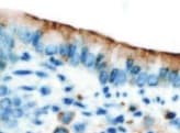
Lipocalin‑2/NGAL in Human Pancreas.
Lipocalin-2/NGAL was detected in immersion fixed paraffin-embedded sections of human pancreas using Rat Anti-Human Lipocalin-2/NGAL Monoclonal Antibody (Catalog # MAB1757) at 15 µg/mL overnight at 4 °C. Tissue was stained using the Anti-Rabbit HRP-DAB Cell & Tissue Staining Kit (brown; Catalog # CTS005) and counterstained with hematoxylin (blue). Specific staining was localized to plasma membrane of ductal cells. View our protocol for Chromogenic IHC Staining of Paraffin-embedded Tissue Sections.
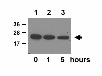
Detection of Human Lipocalin‑2/NGAL by Western Blot.
Western blot shows lysates of Capan-1 human pancreatic adenocarcinoma cell line, HT-29 human colon adenocarcinoma cell line, and human bone marrow tissue. PVDF membrane was probed with 0.2 µg/mL of Rat Anti-Human Lipocalin-2/NGAL Monoclonal Antibody (Catalog # MAB1757) followed by HRP-conjugated Anti-Rat IgG Secondary Antibody (Catalog # HAF005). A specific band was detected for Lipocalin-2/NGAL at approximately 22 kDa (as indicated). This experiment was conducted under reducing conditions and using Immunoblot Buffer Group 1.
Detection of Lipocalin-2/NGAL by Western Blot
Silencing of LCN2 impairs cell cycle‐associated proteins. (A) The top proteins downregulated in LCN2‐silenced cells compared with control cells after RPPA proteomic analysis. (B) Gene set enrichment analysis of RPPA data identified pathways that are enriched or downregulated in control vs LCN2‐silenced SUM149 cells. (C) STRING interaction network of predicted active kinases based on enrichment of kinase substrates and protein interactions identified using KEA. The confidence of the interaction is reflected by the edge thickness. Based on node distribution analysis, four central proteins were identified (MAPK1, MAPK8, RPS6KB1, and MTOR). (D) Silencing of LCN2 in SUM149 cells reduced pMEK and pERK expression. Image collected and cropped by CiteAb from the following open publication (https://pubmed.ncbi.nlm.nih.gov/34342930), licensed under a CC-BY license. Not internally tested by R&D Systems.
Detection of Lipocalin-2/NGAL by Western Blot
LCN2 was highly expressed in tumors from patients with IBC. (A) High LCN2 expression was associated with shorter overall survival in a meta‐dataset of patients with non‐IBC. (B, C) LCN2 mRNA expression was higher in tumors from IBC patients vs non‐IBC patients in two independent breast cancer datasets [IBC World Consortium Dataset; GSE45582]. (D) LCN2 mRNA expression was higher in ER‐negative compared to ER+ samples IBC samples. (E) LCN2 mRNA expression was higher in more aggressive molecular subtypes, ERBB2+ and TNBC, compared to HR‐positive/HERBB2‐negative subtype. (F) LCN2‐high expression correlates with shorter overall survival in patients with IBC. (G) LCN2 mRNA expression was higher in IBC cell lines compared to non‐IBC cell lines. (H, I) LCN2 protein expression was higher in IBC cell lines compared to non‐IBC cell lines shown by (H) immunoblotting or (I) ELISA for secreted LCN2 in supernatants. Bar graphs indicate mean ± SEM from three independent experiments. graphpad prism software was used to obtain the P values, with Mann–Whitney tests used to compare two categories or one‐way analysis of variance to compare three or more categories. Black lines in each group (B–E, and G) indicate mean ± SD. Image collected and cropped by CiteAb from the following open publication (https://pubmed.ncbi.nlm.nih.gov/34342930), licensed under a CC-BY license. Not internally tested by R&D Systems.Applications for Human Lipocalin‑2/NGAL Antibody
Application
Recommended Usage
Immunohistochemistry
8-25 µg/mL
Sample: Immersion fixed paraffin-embedded sections of human pancreas
Sample: Immersion fixed paraffin-embedded sections of human pancreas
Western Blot
0.2-1 µg/mL
Sample: K562 human chronic myelogenous leukemia cell line, Capan‑1 human pancreatic adenocarcinoma cell line, HT‑29 human colon adenocarcinoma cell line, and human bone marrow tissue
Sample: K562 human chronic myelogenous leukemia cell line, Capan‑1 human pancreatic adenocarcinoma cell line, HT‑29 human colon adenocarcinoma cell line, and human bone marrow tissue
Reviewed Applications
Read 2 reviews rated 5 using MAB1757 in the following applications:
Formulation, Preparation, and Storage
Purification
Protein A or G purified from hybridoma culture supernatant
Reconstitution
Reconstitute at 0.5 mg/mL in sterile PBS. For liquid material, refer to CoA for concentration.
Loading...
Formulation
Lyophilized from a 0.2 μm filtered solution in PBS with Trehalose. *Small pack size (SP) is supplied either lyophilized or as a 0.2 µm filtered solution in PBS.
Shipping
Lyophilized product is shipped at ambient temperature. Liquid small pack size (-SP) is shipped with polar packs. Upon receipt, store immediately at the temperature recommended below.
Stability & Storage
Use a manual defrost freezer and avoid repeated freeze-thaw cycles.
- 12 months from date of receipt, -20 to -70 °C as supplied.
- 1 month, 2 to 8 °C under sterile conditions after reconstitution.
- 6 months, -20 to -70 °C under sterile conditions after reconstitution.
Calculators
Background: Lipocalin-2/NGAL
References
- Flower, D.R. et al. (1994) FEBS Lett. 354:7.
- Kjeldsen, L. et al. (1993) J. Biol. Chem. 268:10426.
- Kjeldsen L, et al. (2000) Biochim Biophys Acta. 1482:272.
- Devireddy, L.R. et al. (2001) Science 293:829.
- Yang, M.B. et al. (2002) Mol. Cell. 10:1045.
- Goetz, D.H. et al. (2002) Mol. Cell 10:1033.
- Flo, T.H. et al. (2004) Nature 432:917.
- Hanai, J. et al. (2005) J. Biol. Chem. 280:13641.
Long Name
Neutrophil Gelatinase-associated Lipocalin
Alternate Names
24p3, LCN2, Lipocalin2, MSFI, NGAL, Oncogene 24p3, p25, Siderocalin, Uterocalin
Gene Symbol
LCN2
UniProt
Additional Lipocalin-2/NGAL Products
Product Documents for Human Lipocalin‑2/NGAL Antibody
Certificate of Analysis
To download a Certificate of Analysis, please enter a lot or batch number in the search box below.
Note: Certificate of Analysis not available for kit components.
Product Specific Notices for Human Lipocalin‑2/NGAL Antibody
For research use only
Related Research Areas
Citations for Human Lipocalin‑2/NGAL Antibody
 Powered by Bioz
Customer Reviews for Human Lipocalin‑2/NGAL Antibody (2)
5 out of 5
2 Customer Ratings
Have you used Human Lipocalin‑2/NGAL Antibody?
Submit a review and receive an Amazon gift card!
$25/€18/£15/$25CAN/¥2500 Yen for a review with an image
$10/€7/£6/$10CAN/¥1110 Yen for a review without an image
Submit a review
Customer Images
Showing
1
-
2 of
2 reviews
Showing All
Filter By:
-
Application: ImmunohistochemistrySample Tested: Lung tissueSpecies: HumanVerified Customer | Posted 10/12/2021Formalin-fixed lung sections

-
Application: Western BlotSample Tested: A549 human lung carcinoma cell lineSpecies: HumanVerified Customer | Posted 02/24/2016
There are no reviews that match your criteria.
Protocols
Find general support by application which include: protocols, troubleshooting, illustrated assays, videos and webinars.
- Antigen Retrieval Protocol (PIER)
- Antigen Retrieval for Frozen Sections Protocol
- Appropriate Fixation of IHC/ICC Samples
- Cellular Response to Hypoxia Protocols
- Chromogenic IHC Staining of Formalin-Fixed Paraffin-Embedded (FFPE) Tissue Protocol
- Chromogenic Immunohistochemistry Staining of Frozen Tissue
- ClariTSA™ Fluorophore Kits
- Detection & Visualization of Antibody Binding
- Fluorescent IHC Staining of Frozen Tissue Protocol
- Graphic Protocol for Heat-induced Epitope Retrieval
- Graphic Protocol for the Preparation and Fluorescent IHC Staining of Frozen Tissue Sections
- Graphic Protocol for the Preparation and Fluorescent IHC Staining of Paraffin-embedded Tissue Sections
- Graphic Protocol for the Preparation of Gelatin-coated Slides for Histological Tissue Sections
- IHC Sample Preparation (Frozen sections vs Paraffin)
- Immunofluorescent IHC Staining of Formalin-Fixed Paraffin-Embedded (FFPE) Tissue Protocol
- Immunohistochemistry (IHC) and Immunocytochemistry (ICC) Protocols
- Immunohistochemistry Frozen Troubleshooting
- Immunohistochemistry Paraffin Troubleshooting
- Preparing Samples for IHC/ICC Experiments
- Preventing Non-Specific Staining (Non-Specific Binding)
- Primary Antibody Selection & Optimization
- Protocol for Heat-Induced Epitope Retrieval (HIER)
- Protocol for Making a 4% Formaldehyde Solution in PBS
- Protocol for VisUCyte™ HRP Polymer Detection Reagent
- Protocol for the Preparation & Fixation of Cells on Coverslips
- Protocol for the Preparation and Chromogenic IHC Staining of Frozen Tissue Sections
- Protocol for the Preparation and Chromogenic IHC Staining of Frozen Tissue Sections - Graphic
- Protocol for the Preparation and Chromogenic IHC Staining of Paraffin-embedded Tissue Sections
- Protocol for the Preparation and Chromogenic IHC Staining of Paraffin-embedded Tissue Sections - Graphic
- Protocol for the Preparation and Fluorescent IHC Staining of Frozen Tissue Sections
- Protocol for the Preparation and Fluorescent IHC Staining of Paraffin-embedded Tissue Sections
- Protocol for the Preparation of Gelatin-coated Slides for Histological Tissue Sections
- R&D Systems Quality Control Western Blot Protocol
- TUNEL and Active Caspase-3 Detection by IHC/ICC Protocol
- The Importance of IHC/ICC Controls
- Troubleshooting Guide: Immunohistochemistry
- Troubleshooting Guide: Western Blot Figures
- Western Blot Conditions
- Western Blot Protocol
- Western Blot Protocol for Cell Lysates
- Western Blot Troubleshooting
- Western Blot Troubleshooting Guide
- View all Protocols, Troubleshooting, Illustrated assays and Webinars
Loading...